Our mission to Mars is driving a need to better understand the physiological impact on the human body. Deep space travel places unique challenges for humans. It is important that there is minimal impact on space travelers' senses. This includes hearing. This study uses our Shank 1a Antibody to determine changes in the neuronal structure of the ear in microgravity.
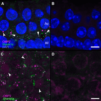
Top Image: Verification of antibodies to CtBP2 and Shank1a. A and B: serial 14-µm cryosections were obtained from a postnatal day 71 (P71) mouse utricle, and maximum-intensity projections are shown. Hair cell (hc) and support cell (sc) nuclei are illuminated by the DAPI stain (blue). Numerous closely associated CtBP2-and Shank1a-positive puncta can be observed in the positive immunostained section represented in A (block arrowheads). The CtBP2-positive puncta highlighted by the flared arrowhead in A may represent an undocked synaptic ribbon. No primary antibodies were included in the processing represented in the micrograph in B. The scale bar in B represents 5 µm and also applies to A. C and D: maximum-intensity projection micrographs from right and left whole mount utricles from a P65 mouse...

Importantly, fixative administration into the temporal bones yielding the specimens represented in C and D was delayed 7 min to replicate the conditions associated with specimens derived from the microgravity and control specimens. The positive-immunostained specimen of the pair is shown in C, where numerous closely associated CtBP2-positive and Shank1a-positive puncta can be observed. Though faint, CtBP2-immunostained nuclei are highlighted by the flared arrowheads. The micrograph in D illustrates the results of withholding primary antibodies from the processing. Immunolabeled puncta are not observed. The scale bar in D represents 5 µm and also applies to C. https://www.physiology.org/doi/full/10.1152/jn.00240.2016
These results demonstrate that structural plasticity was topographically localized to the utricular region that encodes very low frequency and static changes in linear acceleration, and illuminates the remarkable capabilities of utricular hair cells for synaptic plasticity in adapting to novel gravitational environments.
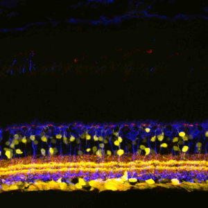
Bottom Image: HC Staining in Mouse Retina, Courtesy of Sal Stella, UCLA


