Just how important is the tumor microenvironment (TME) in cancer research? As a supplier of primary human cancer-associated fibroblasts (CAFs), we’re exposed to plenty of studies looking into their role. One big theme: it’s becoming ever clearer that cancer treatments must take CAFs and their role in the TME into account.
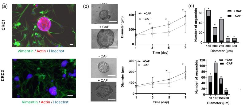
Image: 3D organotypic colorectal tumor model surrounded by CAFs (vimentin, green) (a). Organoids are larger and more plentiful when CAFs are incorporated than without CAFs (b & c).
In a newly released paper, a team of researchers from the University of Akron builds 3D organoids with patient-derived colorectal tumor samples to study prospective cancer treatments. They compare how organoids develop, prosper, and react to treatments when they are with and without colorectal tumor CAFs (cat. CAF115) provided by Neuromics.

Some of the findings:
- When CAFs are mixed with colorectal tumor cells, they promote larger and an increased number of tumor organoids.
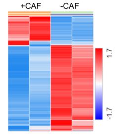
- Gene expression in tumor cells changes dramatically when mixed with CAFs, as CAFs activate various pathways.
- Hepatocyte growth factor (HGF) secreted by CAFs activates MET receptors in patient-derived tumor cells. The activation promotes proliferation and stemness in cancer cells.
- The presence of CAFs in the organoids made treatments less effective, as CAFs enable drug resistance in cancer.
- This tumor model has the potential to be used with individualized patient samples for custom treatments.
Check out the full paper here: https://aacrjournals.org/mct/article/doi/10.1158/1535-7163.MCT-24-0756/760710
Bottom Image: Heatmap of gene expression in colorectal tumor organoids shows changes with or without CAFs. Each row represents a gene, each column a sample, and every cell shows normalized gene expression values.
This study is just one of several recent papers utilizing Neuromics CAFs to better understand the TME. Australian researchers used our pancreatic CAFs (cat. CAF118) to see how they react to small extracellular vesicles released by pancreatic cancer cells (learn more). Collaborating investigators from Takeda, BioTuring, and BMS also employed our Pancreatic CAFs in 2D and 3D monocultures and cocultures. They found that the expression profile and pro-proliferative effects of CAFs vary substantially depending on the culture conditions (check out our blog post).
You can explore all our human CAF options here: https://www.neuromics.com/fibroblasts-cafs-cancer-cells


