Far and away, the most common research theme with our human cancer associated fibroblasts (CAFs) is 3D. Whether it's 3D co-culture, organoids, or in vivo models, the value of these cells is most apparent when integrated into 3D models that mimic the tumor microenvironment (TME). A new study from scientists at the Federal University of Santa Catarina in Brazil outlines a novel approach for building a 3D platform for co-culturing cells. The investigators utilized our human primary breast CAFs (cat. CAF116) and US origin premium fetal bovine serum (FBS) (cat. FBS001) to make it happen.
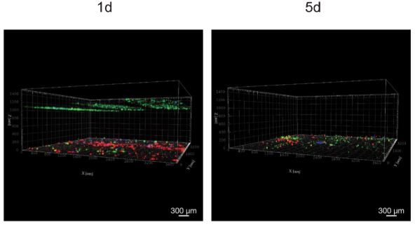
Image: 3D confocal microscopy of cells seeded into BNC at different time points: MDA-MB-231 cells (green), Neuromics breast CAF (red), and M2 macrophages (blue). MDA-MB-231 cells and macrophages were grown with Neuromics’ FBS.
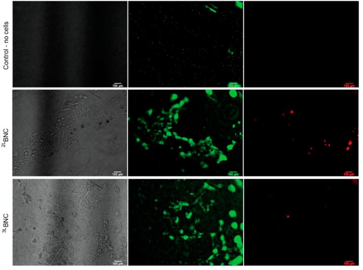
The investigators outlined the steps to build a translucent multi-compartmentalized stacked multilayered nanocellulose scaffold. The scaffold consisted of bacterial nanocellulose (BNC) layers separated by interlayers of a lower density of nanocellulose fibers. To validate their scaffold model, they cultured several cell types, including EOMA cells, EA.hy926 cells, MDA-MB-231 cells, monocytes, and macrophages with media supplemented with Neuromics' FBS.

Then, the researchers injected MDA-MB-231 cells, M2 macrophages, and our breast CAFs into a triple-layered BNC scaffold to mimic the TME. The BNC scaffold was validated as a reliable 3D co-culture model by performing various assays. Cells remained viable for 15 days, breast cancer-related genes expressed as expected, and the cells interacted as expected. You can check out the complete study here.
Bottom Image: EOMA cells grown with Neuromics’ FBS after one-week cell culture in BNC scaffolds.
Our CAFs and FBS are two of our most commonly cited product categories. Explore all publications using these products here. Furthermore, check out all our human CAF options here and FBS options here.


